Hospital imaging equipment is giving researchers a new, noninvasive window into ancient Egyptian remains — and the results are changing how museums study and preserve fragile collections. Using advanced CT technology, teams in Budapest are uncovering medical details and burial practices from millennia ago without unwrapping a single bandage, a development that matters now for conservation, scholarship, and public display.
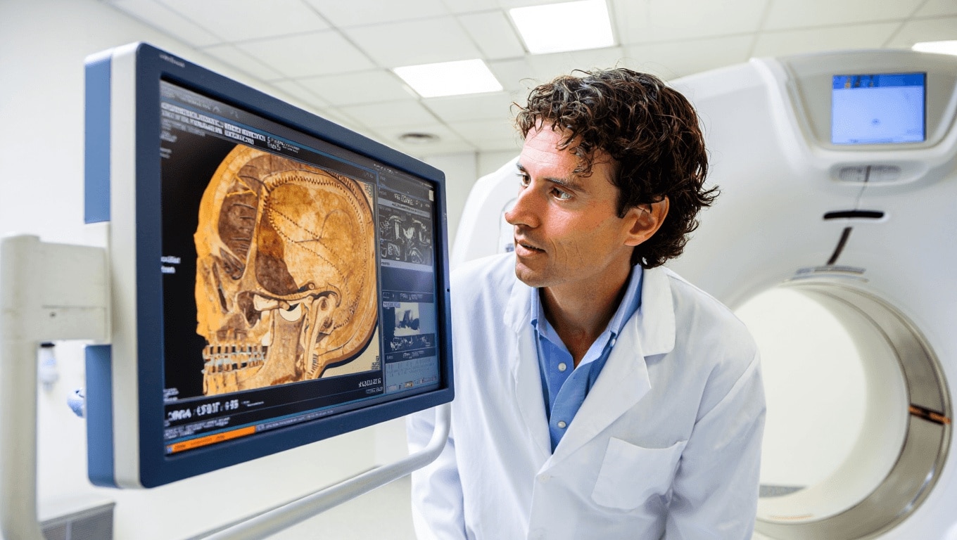
At Semmelweis University’s Medical Imaging Center, scientists are running high-end hospital scanners outside regular patient hours to examine mummies from museum collections. The machines — clinical-grade CT scanners outfitted with the latest photon-counting detectors — produce extremely detailed two- and three-dimensional images that reveal internal structures and materials that were previously inaccessible without destructive sampling.
Staff emphasized scans were performed at night so routine patient services wouldn’t be interrupted. The detail of that scheduling is striking: it underscores how museums and hospitals are now sharing resources to balance public health needs with research priorities.
What the scans can show
These scans are doing more than satisfying curiosity. They provide concrete, testable information about the lives and care of people from antiquity, and they inform how collections are handled today.
- Health and disease: Bone density and joint conditions such as osteoporosis and arthritis, dental problems like tooth decay, and other pathological signs can be identified from images alone.
- Biological profile: Cranial and skeletal measurements help estimate age at death and can flag developmental anomalies that point to childhood health or nutritional stress.
- Mummification techniques: Layering of linen, packing materials and hidden objects — including metal or stone amulets — are visible between wrappings without physically opening the bundle.
- Object identification: Items or body parts that seemed ambiguous from the outside can be clearly identified; in one case a wrapped bundle thought to be a head or a bird was revealed to be a human foot.
- Conservation planning: Imaging maps internal fragility, guiding curators on how to stabilize and transport artifacts safely.
Radiology-level imaging makes it possible to revisit old assumptions. Specimens that were once too delicate for study can now be assessed repeatedly over time, enabling longitudinal monitoring of conservation interventions.
Beyond individual finds, the technology is widening the lens on ancient health. By compiling scan data across many mummies, researchers can start to chart patterns of disease, injury, and mortuary practice across regions and centuries — a kind of population-level paleopathology that doesn’t require physical sampling.
Why this matters now
Two advances are driving the moment: the availability of clinical scanners to research partners and the emergence of photon-counting detector technology, which improves contrast and material discrimination at lower radiation doses. That combination increases what can be seen inside a specimen while reducing risk to the object.
The implications are practical and ethical. Museums can minimize invasive conservation work, researchers can generate publishable data without destructive tests, and descendant communities gain a less intrusive way to study ancestral remains. At the same time, hospitals and cultural institutions must negotiate scheduling, privacy and chain-of-custody issues when using medical facilities for research scans.
For museums, the result is a shift in how collections are managed: from boxes of fragile objects to repositories of rich, image-based data that can be re-analyzed as methods improve. For historians and medical researchers, it’s an expanding archive of direct evidence about ancient lifeways and health.
As scanning becomes more widespread, curators and scientists will face new questions about access, data sharing and the ethics of digitally intrusive research on human remains. The current work in Budapest illustrates both the scientific promise and the logistical trade-offs of this approach — and signals that noninvasive imaging will be central to archaeological science going forward.
Similar Posts
- Rarest mineral confirmed: a single 0.011 oz specimen reveals gaps in Earth’s catalog
- Shark fossils in Arkansas turn the state into a skeleton hotspot
- Ancient Vulture Nests Reveal 750-Year-Old Shoe: Discover Hidden Human Artifacts!
- Oldest animal status challenged by new study: iconic fossils may not be animals
- Memory loss in aging: brain changes that make remembering harder

Miles Harper focuses on optimizing your daily life. He shares practical strategies to improve your time management, well-being, and consumption habits, turning your routine into lasting success.